MedPharmRes
University of Medicine and Pharmacy at Ho Chi Minh City
Original article

ADAMTSL3 - a potential direct target of miR-144-3p in chondrocyte cell

Phuong Thi Bich Hoahttps://orcid.org/0000-0003-4732-4558, Quan Dang Nguyenbhttps://orcid.org/0000-0002-4333-2086, Tram Thi Kim Phambhttps://orcid.org/0000-0002-6840-3338, Anh Ha Nam Tangc, Linh Thi Truc Lea,*https://orcid.org/0000-0003-3879-2181
aBiotechnology Department , Ho Chi Minh City Open University, Ho Chi Minh City, Vietnam
bBiotechnology Center of Ho Chi Minh City, Ho Chi Minh City, Vietnam
cNguyen Tri Phuong Hospital, Ho Chi Minh City, Vietnam
*Address correspondence: to Linh Thi Truc Le at the Biotechnology Department , Ho Chi Minh City Open University, Ho Chi Minh City, Vietnam; E-mail: linh.ltt@ou.edu.vn

© Copyright 2023 MedPharmRes. This is an Open-Access article distributed under the terms of the Creative Commons Attribution Non-Commercial License (http://creativecommons.org/licenses/by-nc/4.0/) which permits unrestricted non-commercial use, distribution, and reproduction in any medium, provided the original work is properly cited.

Received: Jul 27, 2022; Revised: Sep 20, 2022; Accepted: Oct 06, 2022

Published Online: Dec 31, 2023

Abstract:

The miR-144-3p was discovered to upregulate in osteoarthritis. However, the function of this molecule is still poorly understood. Identification of microRNA target genes could shed a light on miRNA functions. In this project, we aim to identify a direct target of miR-144-3p. Potential targets of miR-144-3p were predicted by bioinformatic algorithms. Expression of ADAMTSL3 in miRNA-144-3p gain-and- loss- of function experiment was quantified by Realtime-PCR. Next, the 3’UTR of ADAMTSL3 containing miR-144-3p-binding sites was subcloned in the 3’;UTR of the Firefly luciferase gene on the pmiRGlo. To test the complementary binding of miR-144-3p to ADAMTSL3 mRNA, we employed the luciferase assay using pmiRGlo-ADAMTSL3 vector or pmiRGlo-ADAMTSL3 mutant in which miR-144-3p binding sites in the 3’UTR was mutated. Results showed that miRNA-144-3p directly inhibits the expression of ADAMTSL3 through its ability to pair at “seed” sites. This study provides evidence for the interaction of miRNA-144-3p and its target, ADAMTSL3 mRNA, in chondrocytes.

Keywords: miR-144-3p; osteoarthritis; target gene; ADAMTSL3

1. INTRODUCTION

Osteoarthritis (OA) is common in musculoskeletal diseases. OA occurs more commonly in the elderly. Approximately 303 million people worldwide suffered from OA in 2017 [1]. By 2050, more than 20% of the world’s population will be over the age of 60, 15% of whom will have symptoms of OA and more than a third of these will be disabled [2]. OA is quite popular in Vietnam but there are no exact statistics. Vietnam’s population aging rate is currently the fastest in Asia. It is predicted that by 2050, Vietnamese people aged 60 and over will be doubled from 11.9 to 29 million, accounting for about a third of the total population. Therefore, Vietnam will witness an increase in OA in the following years because OA is proportional to population aging.

MicroRNA (miRNA) is an RNA molecule with about 20-25 nucleotides that is able to regulate gene expression. Binding of miRNAs to its specific binding site on the 3’UTR region of the mRNA leads to mRNA hydrolysis or the inhibition of translation [3, 4]. Aberrant expression of miRNAs is reported in many diseases such as cancer and metabolic disorders [5]. Abnormal expression of several OA-associated miRNAs has been identified such as miR-483, miR-22, miR-9, miR-98, miR-485-5p, miR-455, miR-210, miR-27b, miR-140, and miR-29 [6-14].

miR-144-3p has been shown to be associated with OA. Specifically, miR-144-3p increased in OA chondrocytes [15] and 5-fold in OA osteoblasts [8]. Moreover, miR-144-3p inhibits CRAT which inhibits osteoclastogenesis [16]. Several genes involved in OA have also been shown to be the targets of miR-144-3p such as Smad4, FZD4 and TET2 [17-19]. The findings suggest that miR-144-3p plays a role in the pathogenesis of OA. However, the function of this miRNA molecule has not been completely revealed. To understand the function of miR-144-3p in OA, it is necessary to identify its target genes. Therefore, we used bioinformatics tools to predict the target genes of miR-144-3p. Next, we demonstrated that this gene was a direct target of miRNA-144-3p in vitro.

2. MATERIALS AND METHOD

2.1. Prediction miR-144-3p potential target genes

miRmap (https://mirmap.ezlab.org/app/), MicroRNA Target Prediction Database - miRDB (http://mirdb.org/), and TargetScanHuman (http://www.targetscan.org/vert_72/) were utilized to predict miR-144-3p potential targets. The sequences of mature hsa-miR-144-3p (miR-144-3p) were obtained from the miRBase database. The sequence of miR-144-3p binding sites were identified as “TACTGT” (6 mer), “ATACTGT” (7 mer-m8), “TACTGTA” (7 mer-A1) and “ATACTGTA” (8 mer).

2.2. Gain - loss function assay

SW1353 chondrocytes were cultured in DMEM media (Thermo Fisher) supplemented with 100 μg/ml of penicillin and streptomycin (Thermo Fisher), and 10% (v/v) fetal bovine serum (Thermo Fisher), at 37°C, 5% (v/v) CO2 until the cell growth reached 80% confluency. The cells were then seeded at a density of 2x10⁵ cells/mL in a 6-well plate and cultured to 80% confluency. Cells were then transfected with 50 nM miR-144-3p mimics, miR-144-3p inhibitor (Qiagen) or corresponding control (Qiagen) using Lipofectamine 2000 (Thermo Fisher) for approximately 24 hours according to the manufacturer’s protocol.

2.3. Quantification by Realtime-PCR

Total RNA from cells was extracted using TRIzol Reagent (Invitrogen) and converted to cDNA using the SuperScript II RT kit (Thermo Fisher). The concentration and purity of extracted total RNA were checked by Nanodrop using A260/280 and A260/230 ratios. Expression of ADAMTSL3 was determined using the SensiFAST SYBR No-ROX Kit (Bioline), forward primer 5’-TGGAAGCATGTGATGAGAGC-3’ and reverse primer 5’-TTGACTGTCTGCTGGGTCTG-3’ with an internal control, 18S rRNA. Expression of ADAMTSL3 was determined using the ΔCT formula. Realtime-PCR quantification was performed using a LightCycler® 96 System (Roche).

2.4. Luciferase reporter assays

The 3’UTR fragment with the miR-144-3p-binding sites of ADAMTSL3 was cloned into pmiRGlo vector by the In-Fusion HD Cloning kit (Takara). The forward primer 5’-GCTCGCTAGCCTCGATTAGGTCAGGGTTGGATTGG-3 ’ and reverse primer 5’-CGACTCTAGACTCGAGGATCATGAAACAGAAACAT TAACTC-3’. The mutated pmiRGlo-ADAMTSL3 vector was kindly provided by an overseas research group. The cloning product was checked by colony PCR using forward primer 5’-TTCACTGACACTGGGTTGAGCACTACTG-3’ and reverse primer 5’-CGACTCTAGACTCGAGGATCATGAAACAGAAACATTAACTC-3’, and subsequently sequenced.

SW1353 cells were seeded in a 96-well plate at a density of 5x10⁴ cells/mL in 100 μL of medium and cultured overnight. The plasmids including pmiRGlo-ADAMTSL3 (100 ng), mutant pmiRGlo-ADAMTSL3 (100 ng), miR-144-3p mimics (50 nM ) and a control (50 nM ) were then transfected seperately using Lipofectamine 2000 (Thermo Fisher). The level of luciferase activities was determined using the Dual-luciferase Reporter kit (Promega) and detected using an EnVision 2103 Multilable (Perkin Elmer). Normalized luciferase activity was reported as the Firefly luciferase/Renilla luciferse activity.

2.5. Statistical analysis

The unpaired two-tailed student’s test was performed to analyze all obtained data using GraphPad Prism version 6.

3. RESULTS

Two binding sites for miR-144-3p on ADAMTSL3 3’UTRs

Among potential target genes of miR-144-3p predicted by bioinformatics algorithms , we selected candidates that have been reported to be associated with OA in previous studies. TargetScanHuman and miRDB predicted that miR-144-3p had round 1043 and 1254 target genes, respectively. The difference in the number of target genes proposed by two bioinformatics tools is probably due to the discrepancy in predictive analytics algorithms.

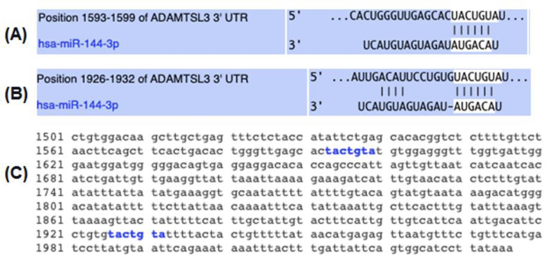
Of these predicted target genes, ADAMTSL3 was identified by both softwares with two seed sites of miR-144-3p on the 3’UTR (Figure 1). TargetScan results showed that ADAMTSL3 has two 7mer-A1 (TACTGTA) binding sites for miR-144-3p, of which position 1593-1599 is highly conserved (Figure 1A) and position 1926-1932 is poorly conserved (Figure 1B). Similarly, the miRDB tool also revealed two 7mer-A1 (TACTGTA) sites for miR-144-3p on the ADAMTSL3 3’UTR were located at position 1593 and 1926 (Figure 1C). These data proposed that miR-144-3p possibly binds to the 3’UTR of ADAMTSL3.

mpr-7-4-41-g1
Figure 1. The 3’UTR of ADAMTSL3 possesses two recognition sites of miR-144-3p. Targetscan predicted two miR-144-3p seed sites on the 3’UTR of ADAMTSL3 with (A) conserved 7mer-A1 site at 1593-1599 and (B) nonconserved 7mer-A1 site at position 1926-1932. miRDB suggested two binding sites of miR-144-3p at positions 1593 and 1926. Seed sites are highlighted in blue (C).
Download Original Figure
ADAMTSL3 expression level is negatively correlated to miR-144-3p abundance in chondrocytes

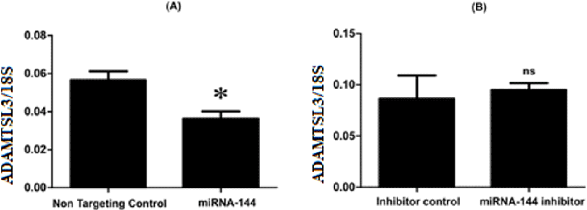
The expressions of ADAMTSL3 in SW1353 cells after being transfected with miR-144-3p mimics or miRNA-144-3p inhibitor and control were observed using Realtime-PCR. Upon miR-144-3p overexpression, there is a reduce in ADAMTSL3 expression compared to non-targeting control (Figure 2A). Meanwhile, the inhibition of miRNA-144-3p had insignificant effect on the expression level of ADAMTSL3 compared to control (Figure 2B).

mpr-7-4-41-g2
Figure 2. The negative correlation between ADAMTSL3 and miRNA-144-3p in cells. (A) The overexpression of miR-144-3p for 24 hours before the quantification of ADAMTSL3 level using RealTime-PCR. (B) Similar to A but inhibiting miRNA-144-3p by using an miR-144-3p inhibitor. 18S was used as an internal control. Data were analyzed using Student’s unpaired t-test, n=3. *p < 0.05.
Download Original Figure
Direct interaction between miR-144-3p and ADAMTSL3

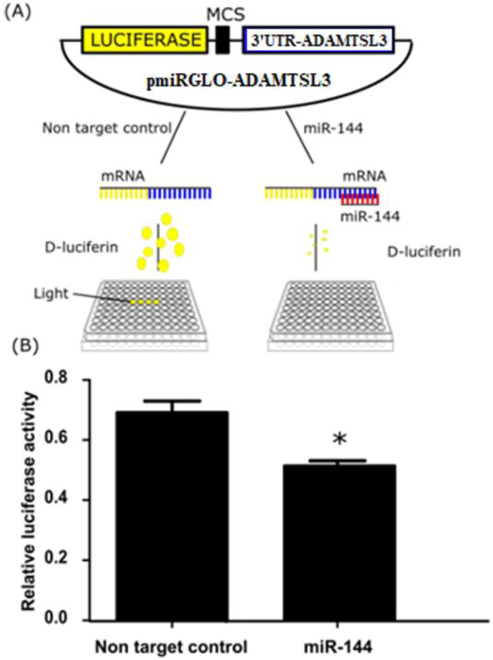
To investigate whether miR-144-3p directly interacts with ADAMTSL3 at the binding sites, the 3’UTR of ADAMTSL3 containing the miR-144-3p-binding was inserted downstream of the Firefly luciferase reporter gene on the pmiRGlo vector. The pmiRGlo-ADAMTSL3 was co-transfected with miR-144-3p mimics into SW1353 cells which were subsequently cultured for 24 hours before measuring luciferase luminescence expression. As expected, the luciferase activity reduced compared with non-target control (Figure 3).

mpr-7-4-41-g3
Figure 3. miR-144-3p inhibits luciferase expression of the Firefly luciferase gene of vector pmiRGlo-ADAMTSL3. (A) The principle of luciferase reporter assay. The 3’UTR with miRNA-144-3p-binding site of ADAMTSL3 was subcloned in 3’UTR of Firefly luciferase gene on pmirGLo. The pmiRGLo-ADAMTSL3 vector together with miR-144-3p mimics were transfected into SW1353 cells. In the cell, miR-144-3p mimics will complementarily bind to seed sites on the pmiRGLo-ADAMTSL3 vector, leading to mRNA degradation or translation inhibition of luciferase activity. (B) The luciferase activity was detected via luminescence miR-144-3p repressed the luciferase activity. Renilla luciferase was used as an interval control. Data were analyzed using Student’s unpaired t-test, n=3, ***p < 0.001.
Download Original Figure

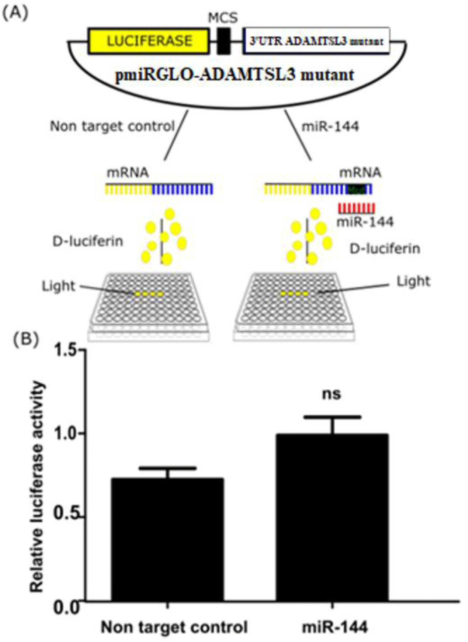
Next, a similar experiment was conducted using mutant pmiRGlo-ADAMTSL3 harboring the two mutants at seed binding sites of miR-144-3p. The mutant vector (100ng) was co-transfected with miR-144-3p mimics or non targeting control for 24 hours and again measured luciferase activity. The relative luciferase activity was recovered compared with controls (Figure 4).

mpr-7-4-41-g4
Figure 4. miR-144-3p did not inhibit the luciferase expression of mutated pmiRGlo-ADAMTSL3 vector. (A) The principle of performing the experiment. The pmiRGlo-ADAMTSL3 vector harbored mutation in miRNA-144-3p-binding sites. The mutant pmiRGLo-ADAMTSL3 vector was co-transfected with miR-144-3p mimics into SW1353 cells. In the cell, miR-144-3p will not be able to complementarily pair with mutated binding site, resulting in a rescue of luciferase expression. The luciferase activity detected via luminescence (B) miR-144-3p did not interfere the luciferase activity of the mutated pmiRGlo-ADAMTSL3 vector. Renilla luciferase was used as an interval control. Data were analyzed using Student’s unpaired t-test, n=4, ***p < 0.001.
Download Original Figure

4. DISCUSSION

We previously identified miR-144-3p overexpression in OA disease [15]. We next determined the function of this molecule by identifying its direct targets by employing various bioinformatics tools and then validating the prediction in vitro. The results indicated that ADAMTSL3 is the direct target of miR-144-3p.

The data of the in silico investigation have identified ADAMTSL3 as a potential target gene of miR-144-3p because on the 3’UTR of the mRNA, there are two miR-144-3p binding sites whose functions are related to OA disease [20]. The novel finding demonstrated that ADAMTSL3 is the target gene of miR-144-3p in OA. Moreover, we found that the expression of ADAMTSL3 was affected by the abundance of miR-144-3p in the cells. In particular, upon the accumulation of miR-144-3p, the expression of ADAMTSL3 decreased. In contrast, upon the depletion of miR-144-3p, ADAMTSL3 marginally increased (not statistically significant). Taken together, the data suggest that miR-144-3p inhibits the expression of ADAMTSL3. However, this result could not determine whether miR-144-3p directly or indirectly (via another molecule) regulates ADAMTSL3. It is necessary to conduct further experiments to answer this question. We also observed an increase of ADAMTSL3 when miR-144-3p in the cells was inhibited, but it was not statistically significant because the endogenous miR-144-3p at the time of the investigation was quite low, which may cause the upregulation of ADAMTSL3.

In addition, we found that when binding sites for miR-144-3p was cloned into the 3’UTR region of the luciferase gene, the activity of reporter gene decreased. The activity was, however, restored when we introduced mutation in the binding sites of miR-144-3p. These results indicated that miR-144-3p acts directly on the ADAMTSL3 mRNA through its binding site on the 3’UTR.

Conclusion

ADAMTSL-3 (also known as punctin-2) is a member of the ADAMTSL family together with ADAMTSL-1, -2, and -3. ADAMTSL-3 is a glycoprotein whose molecular weight is 210-kDa. It possesses 13 TS repeats and is present in the extracellular matrix of various tissues [21]. The function of ADAMTSL-3 has not yet been fully defined. It has been reported to involve both in cell-extracellular matrix interactions and cell-cell interactions as well as control the function of ADAMTS proteases [21].

The increased miR-144-3p in OA was observed previously [15], and the current finding suggests that miR-144-3p may have an adverse interaction with ADAMTSL3. The disregulation of miR-144-3p could lead to the abbrent in the interaction between chondrocyte cells and extracellular matrix. This may consequently lead to OA. However, further studies need to be done before clinical applications.

LIST OF ABBREVIATIONS

miRNA:

microRNA

ADAMTSL3:

A Disintegrin-Like And Metalloprotease Domain With Thrombospondin Type I Motifs-Like 3

3’UTR:

3’ unstranslated region

OA:

Osteoarthritis

FUNDING

This study was funded by the Vietnam Ministry of Education and Training, number B2019-MBS-562-10.

CONFLICT OF INTEREST

The authors declare that there is no conflict of interest.

AUTHORS’ CONTRIBUTION

Linh Thi Truc Le designed the study, Phuong Thi Bich Ho, Linh Thi Truc Le did all the experiments. All authors interpreted the data and contributed in writing of the manuscript. All authors approved the final version to be published.

REFERENCES

1.

James SL, Abate D, Abate KH, Abay SM, Abbafati C, Abbasi N, et al. Global, regional, and national incidence, prevalence, and years lived with disability for 354 diseases and injuries for 195 countries and territories, 1990-2017: a systematic analysis for the Global Burden of Disease Study 2017. The Lancet. 2018 Nov; 392(10159):1789-1858.

2.

Lutz W, Sanderson W, Scherbov S. The coming acceleration of global population ageing. Nature. 2008 Feb; 451(7179):716-719

3.

Ambros V. The functions of animal microRNAs. Nature Sep. 2004; 431(7006):350

4.

Bartel DP. MicroRNAs: genomics, biogenesis, mechanism, and function. Cell. 2004 Jan; 116(2):281-297

5.

Saliminejad K, Khorram Khorshid HR, Soleymani Fard S, Ghaffari SH. An overview of microRNAs: biology, functions, therapeutics, and analysis methods. Journal of cellular physiology. 2019 May; 234(5):5451-5465

6.

Díaz-Prado S, Cicione C, Muiños-López E, Hermida-Gómez T, Oreiro N, Fernández-López C, et al. Characterization of microRNA expression profiles in normal and osteoarthritic human chondrocytes. BMC musculoskeletal disorders. 2012 Aug; 13(1):144

7.

Iliopoulos D, Malizos KN, Oikonomou P, Tsezou A. Integrative microRNA and proteomic approaches identify novel osteoarthritis genes and their collaborative metabolic and inflammatory networks. PloS one. 2008 Nov; 3(11)e3740

8.

Jones S, Watkins G, Le Good N, Roberts S, Murphy C, Brockbank S, et al. The identification of differentially expressed microRNA in osteoarthritic tissue that modulate the production of TNF-α and MMP13. Osteoarthritis and cartilage. 2009 Apr; 17(4):464-472

9.

Chen H, Zhang L, Tang Z, Gong Z. MiR-485-5p promotes the development of osteoarthritis by inhibiting cartilage differentiation in BMSCs. Eur Rev Med Pharmacol Sci. 2018 Jun; 22(11):3294-3302.

10.

Hu S, Zhao X, Mao G, Zhang Z, Wen X, Zhang C, et al. MicroRNA-455-3p promotes TGF-β signaling and inhibits osteoarthritis development by directly targeting PAK2. Experimental & molecular medicine. 2019 Oct; 51(10):1-13

11.

Mizuno Y, Tokuzawa Y, Ninomiya Y, Yagi K, Yatsuka-Kanesaki Y, Suda T, et al. miR-210 promotes osteoblastic differentiation through inhibition of AcvR1b. FEBS letters. 2009 Jul; 583(13):2263-2268

12.

Akhtar N, Rasheed Z, Ramamurthy S, Anbazhagan AN, Voss FR, Haqqi TM. MicroRNA-27b regulates the expression of matrix metalloproteinase 13 in human osteoarthritis chondrocytes. Arthritis & Rheumatism. 2010 May; 62(5):1361-1371

13.

Miyaki S, Sato T, Inoue A, Otsuki S, Ito Y, Yokoyama S, et al. MicroRNA-140 plays dual roles in both cartilage development and homeostasis. Genes & development. 2010 Jun; 24(11):1173-1185

14.

Le LT, Swingler TE, Crowe N, Vincent TL, Barter MJ, Donell ST, et al. The microRNA-29 family in cartilage homeostasis and osteoarthritis. Journal of molecular medicine. 2016 May; 94(5):583-596

15.

Le L, Ho P, Clark I. The role of microRNA 144 in osteoarthritis. Osteoarthritis and cartilage. 2018 Apr; 26:S161

16.

Song J, Kang Y-H, Yoon S, Chun C-H, Jin E-J. HIF-1α: CRAT: miR-144-3p axis dysregulation promotes osteoarthritis chondrocyte apoptosis and VLCFA accumulation. Oncotarget. 2017 Sep; 8(41):69351

17.

Huang C, Geng J, Wei X, Zhang R, Jiang S. MiR-144-3p regulates osteogenic differentiation and proliferation of murine mesenchymal stem cells by specifically targeting Smad4. FEBS letters. 2016 Mar; 590(6):795-807

18.

Sun Z, Wu F, Yang Y, Liu F, Mo F, Chen J, et al. MiR-144-3p inhibits BMSC proliferation and osteogenic differentiation via targeting FZD4 in steroid-associated osteonecrosis. Current pharmaceutical design. 2019 Sep; 25(45):4806-4812

19.

Li N, Liu L, Liu Y, Luo S, Song Y, Fang B. miR-144-3p suppresses osteogenic differentiation of BMSCs from patients with aplastic anemia through repression of TET2. Molecular Therapy-Nucleic Acids. 2020 Mar; 19:619-626

20.

Mead TJ, Apte SS. ADAMTS proteins in human disorders. Matrix Biology. 2018 Oct; 71:225-239

21.

Hall NG, Klenotic P, Anand-Apte B, Apte SS. ADAMTSL-3/punctin-2, a novel glycoprotein in extracellular matrix related to the ADAMTS family of metalloproteases. Matrix Biology. 2003 Nov; 22(6):501-510