Case study

Endoscopic submucosal dissection by using clutch cutter and it knife 2 for early gastric cancer with severe submucosal fibrosis: a case report

Le Quang Nhana,*, Mai Vien Phuonga, Le Quang Nghiab
Author Information & Copyright
aEndoscopy Department, University Medical Center at Ho Chi Minh City, Viet Nam
bGeneral Surgery Department, University of Medicine and Pharmacy at Ho Chi Minh City, Viet Nam
*Address correspondence to Le Quang Nhan, Email quangnhan1947@yahoo.com, Endoscopy depatment, University Medical Center at HO Chi Minh city, Viet Nam and General Surgery Department, University of Medicine and Pharmacy at Ho Chi Minh city, Viet Nam

© Copyright 2018 MedPharmRes. This is an Open-Access article distributed under the terms of the Creative Commons Attribution Non-Commercial License (http://creativecommons.org/licenses/by-nc/4.0/) which permits unrestricted non-commercial use, distribution, and reproduction in any medium, provided the original work is properly cited.

Received: Feb 08, 2018; Revised: Jul 23, 2018; Accepted: Aug 24, 2018

Published Online: Sep 30, 2018

Abstract

Introduction:

Gastric cancer is one of the highly malignant gastrointestinal cancer and the third leading cause of cancer death. In last decade, early gastric cancer (EGC) has been reported by using narrow-band imaging (NBI) magnifying endoscopy. Advances in endoscopic techniques, such as endoscopic submucosal dissection (ESD), have enabled the en bloc resection of these EGC. Although ESD is performed for early gastric cancer, there are still many difficult problems in technique of this procedure. The difficulty of gastric ESD depends on the size and location of a tumor, presence of severe submucosal fibrosis, presence of ulceration... We report a case of our successful ESD by using Clutch cutter and IT knife 2 in treatment of EGC with severe submucosal fibrosis.

Case presentation:

A 62 years old man felt an epigastric discomfort for two months. The narrow-band imaging (NBI) magnifying endoscopy revealed a suspected early gastric cancer type 0 - Ila + lie (Japanese classification for early gastrointestinal cancers) at the incisura angularis, the size of this lesion was 15 mm in diameter, and pathological result of endoscopic biopsy was a well differentiated adenocarcinoma. ESD was performed and we found there were severe submucosal fibrosis which were dissected safer and faster by using Clutch cutter and IT knife 2. There were no complications such as severe bleeding and perforation. The size of resected specimen was 60 x 35 mm and the time of procedure was 150 minutes. After ESD, the pathological result was a well differentiated adenocarcinoma, pTla, UL(-), LY(-), V(-), no cancer cell in vertical and horizontal margins. The healing time of ESD-induced ulcer was 5 weeks without local recurrence.

Conclusion:

Through this case, we aim to emphasize the importance of using Clutch cutter and IT knife 2 as a modified technique which makes ESD a safe procedure in treatment of EGC with severe submucosal fibrosis.

Keywords: Early gastric cancer; submucosal fibrosis; Clutch cutter; IT knife 2; endoscopic submucosal dissecion

1. INTRODUCTION

Early gastric cancer (EGC) is defined as a cancer confined to the mucosal or the submucosal layer (T1 cancer), and regardless of whether regional lymph node metastases are present [5], Endoscopic submucosal dissection (ESD) was developed in the late 1990s as an advanced therapeutic endoscopy; and in 2015, ESD has become a standard treatment for early digestive cancers in Japan and in many other countries [4], For treatment of EGC by ESD, Japanese experts advance the expanded criteria: (1) a differentiated intramucosal adenocarcinoma ≤ 2 cm, without ulcer formation; (2) an ulcer ≤ 3 cm in diameter, with differentiated intramucosal adenocarcinoma; (3) a differentiated adenocarcinoma that has invaded the submucosa, size ≤ 3 cm in diameter [3,6], Surgery is chosen for other lesions and advanced gastric cancers [6].

Although ESD is performed for early gastric cancer, there are still many difficult problems in technique of this procedure. The difficulty of gastric ESD depends on the big size and location of a tumor, presence of severe submucosal fibrosis... Therefore, we introduce our first experiences in Viet Nam in using Clutch cutter and IT knife 2 to make ESD a safe procedure in treatment of EGC which has severe submucosal fibrosis.

CASE PRESENTATION

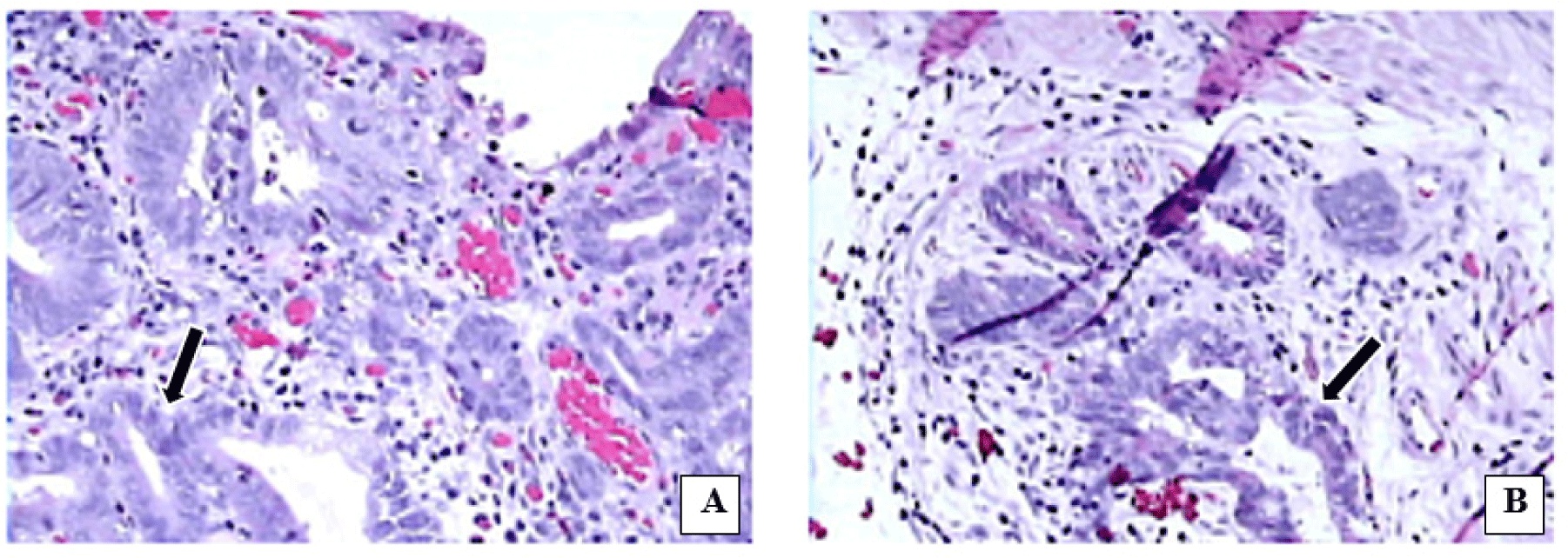
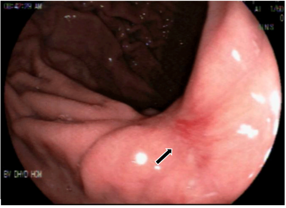
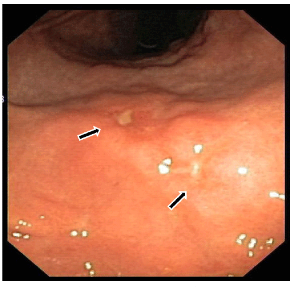
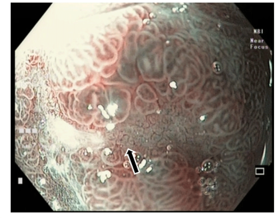
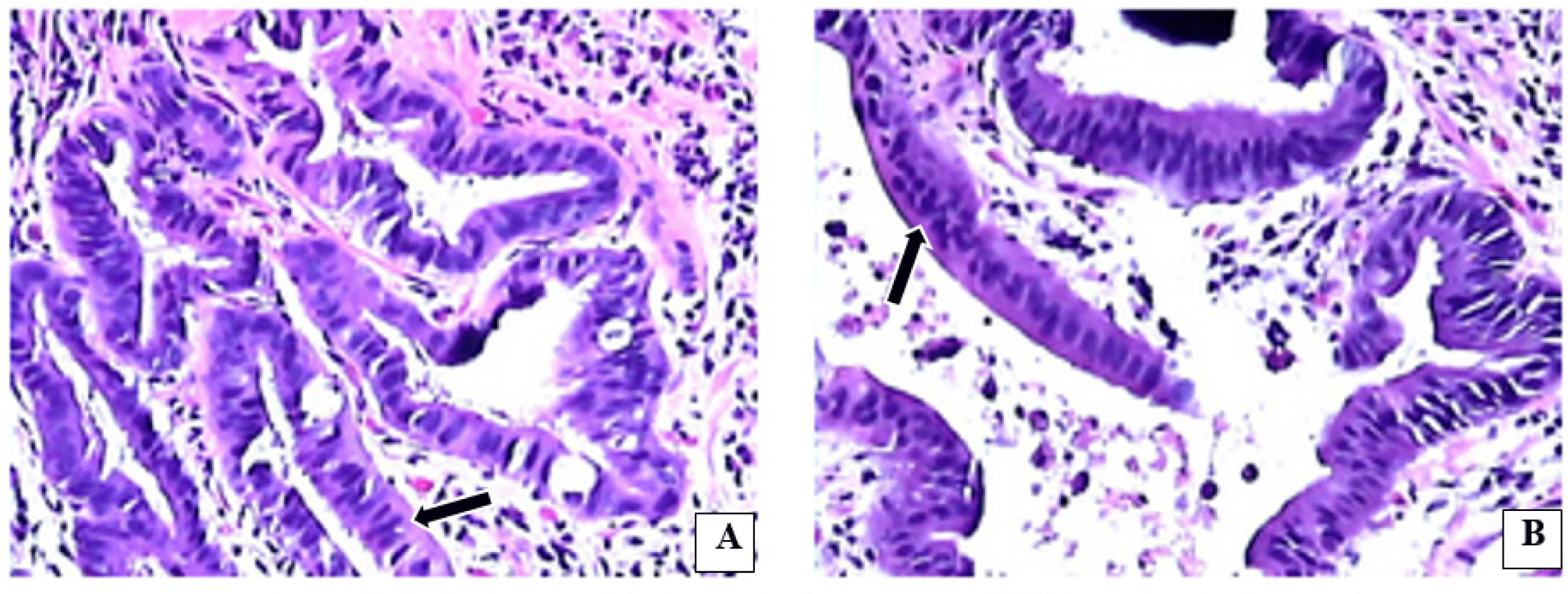
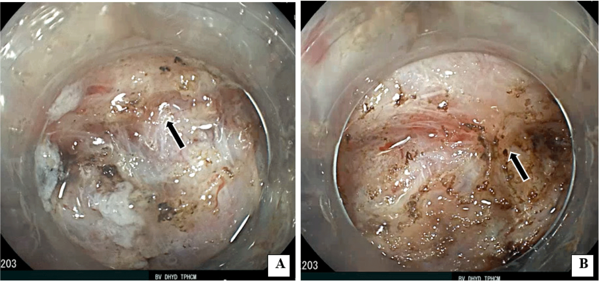
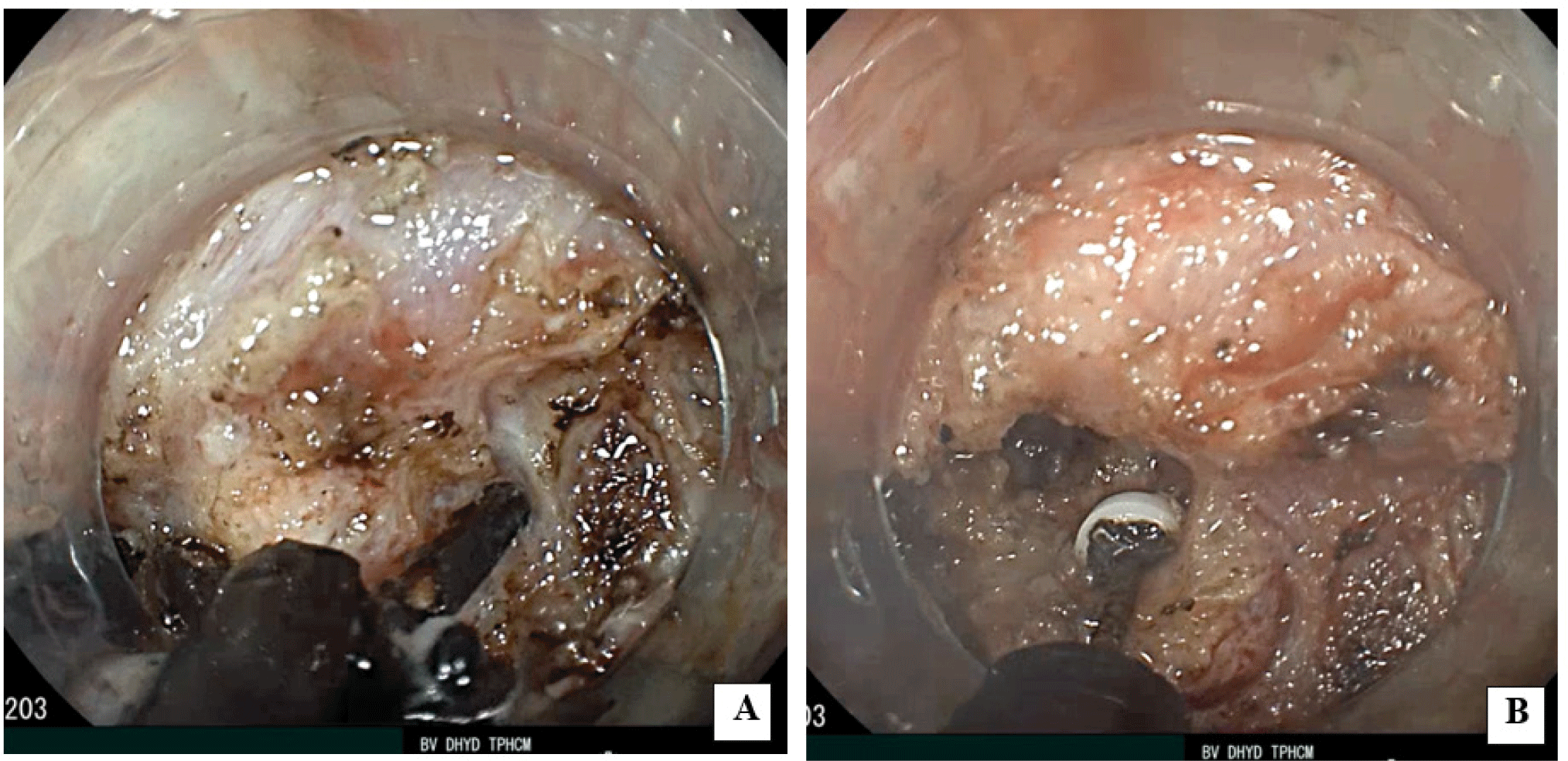
In this study, we describe a case of EGC in a 62-year- old man felt an epigastric discomfort for two months. This patient had no other symptoms. Physical examination showed no abnormalities, and laboratory findings were within normal limits. Under the white light conventional esophagogastroduodenoscopy (EGD), we detected a suspected early gastric cancer type 0 - Ila + Ilc (Japanese classification for early gastrointestinal cancers) and the size of this lesion was 15 mm in diameter. This lesion was located at the incisura angularis (Fig. 1). We found this was a differentiated-type adenocarcinoma with a clear demarcation line, irregular pit pattern and microvascular pattern by using narrow-band imaging (NBI) magnifying endoscopy (Fig. 2). We took one target biopsy and the pathology of this specimen revealed the well-differentiated adenocarcinoma (Fig. 3). We took informed consent before ESD and this patient agreed to this publication. This study was carried out with approval from the ethical committee of University Medical Center at Ho Chi Minh city, Viet Nam.

mpr-2-3-17-g1
Fig. 1. The Lesion was located at the incisura angularis. (⮕).
Download Original Figure
mpr-2-3-17-g2
Fig. 2. NBI magnifying endoscopy showed that this lesion was a differentiated-type adenocarcinoma with a clear demarcation line, an irregular microsurface and microvascular pattern (⮕).
Download Original Figure
mpr-2-3-17-g3
Fig. 3. The pathological result of endoscopic biopsy was well differentiated adenocarcinoma. (a) and (b) There were many cells with an abnormal nuclear(⮕).
Download Original Figure

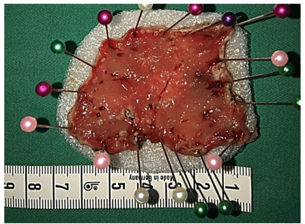
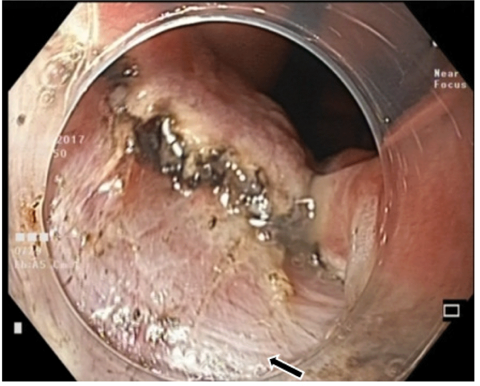
We performed ESD by using a single-channel endoscope (GIF HQ190; Olympus Medical Systems Co., Japan) with an attachment (D-201-11804; Olympus Medical System Co., Japan). To reduce intraluminal gas, a CO2 indufflation pump (Mediavator, USA) was used during the procedure. For this ESD, we used electrosurgical generator which was named VIO300D (ERBE, Tübingen, Germany). We set this generator to the Endo-Cut mode (Effect 3, 60W) for incision of the mucosa and to the Endo-Cut mode (Effect 3, 60W) for dissection of the submucosa. Bleeding was controlled using Coagrasper (FD-411UR; Olympus Medical System Co., Japan) in the soft coagulation mode (50W). A combination of 0.4% sodium hyaluronate (MucoUp; Seikagaku, Tokyo, Japan) and indigo carmine was injected into the submucosal layer to lift the lesion up. Then, we performed circumferential cutting with a dual knife (KD-655L; Olympus Medical System Co, Japan). During the submucosal dissection, we found submucosal white muscular structures which were severe submucosal fibrosis (Fig. 4). We used Clutch cutter (DP2618DT; Fujifilm Co., Japan) and IT knife 2 (KD- 611L; Olympus Medical System Co., Japan) for dissection of these severe submucosal fibrosis (Fig. 5). Finally, ESD was performed sucessfully without perforation and severe bleeding (Fig. 7). We performed en-bloc resection and the size of resected specimen was 60 x 35 mm (Fig. 6). The time of this procedure was 150 minutes.

mpr-2-3-17-g4
Fig. 4. This EGC had severe submucosal fibrosis. (a) There are a little loose submucosal layer which was boundered by severe submucosal fibrosis. (b) The submucosal fibrosis attached severe to the muscular layer (⮕).
Download Original Figure
mpr-2-3-17-g5
Fig. 5. Dissection of the severe submucosal fibrosis. (a) Cluth cutter was used to make shallow holes. (b) We dissected the severe submucosal fibrosis from these shallow holes by using IT knife 2.
Download Original Figure
mpr-2-3-17-g6
Fig. 6. Resected specimen after the ESD. We performed en-bloc resection. The resected specimen was 60x 35 mm
Download Original Figure
mpr-2-3-17-g7
Fig. 7. Endoscopic view after the ESD. The muscular layer could be seen after ESD(⮕)
Download Original Figure

3. DISCUSSION

EGC is a commonly encountered clinical problem in cases undergoing EGD. With the development of digestive endoscopic diagnosis and treatment technology, early digestive cancers can be detected in tiny lesions and treated by endoscopic resection. At present, the best method for endoscopic resection of EGC is ESD [6], ESD has had high en bloc resection rate 93% and high rate of recurrence free at 5 years 100% but the bleeding complication rate of 1.6% and perforation rate of 2.5% [2], The high risk of perforation rate and low rate of en bloc resection caused by EGCs have severe submucosal fibrosis. The severe submucosal fibrosis makes unclear margin between submucosa and muscularis layers, the endoscopitsts will dissect superficially towards to mucosa. This can cause an incomplete resection and high rate of local recurrence. The other harmful effect of severe submucosal fibrosis is high rate of perforation because the endoscopists can dissect deep into the muscularis layers.

The current study showed perforation rates of ESD using a IT knife 2 and Clutch cutter was 1.6% and 3.6%, respectively [1], In this case which was severe submucosal fibrosis, we used both of these devices as a modified technique to finish the ESD without perforation and successful en-bloc resection (Fig. 8). The Clutch cutter was a grasping-type scissor knife and we used it to create the shallow holes. From these shallow holes, we dissected the severe submucosal fibrosis by using IT knife 2. These steps were performed carefully to avoid perforation.

mpr-2-3-17-g8
Fig. 8. The pathological result of resected specimen. After ESD, the pathological result was a well differentiated adenocarcinoma, pTla, UL(-), LY(-),V(-), no cancer cell in lateral and vertical margins. There were many cells with abnormal nuclears(⬅)
Download Original Figure
mpr-2-3-17-g9
Fig. 9. ESD-induced ulcer healed after 5 weeks. There was a scar at the incisura angularis (⮕)
Download Original Figure

4. CONCLUSION

Through this case, we aim to emphasize the importance of using Clutch cutter and IT knife 2 as a modified technique which makes ESD a safe procedure in treatment of EGC with severe submucosal fibrosis.

ACKNOWLEDGEMENTS

Not applicable

FUNDING

The authors declare that they have no funding source

AVAILABILITY OF DATA AND MATERIALS

Not applicable

AUTHORS’ CONTRIBUTION

All authors have read and approved the final manuscript. All authors (NLQ, PMV,NQL) helped on the diagnosis and treatment, data collection and scientific writing.

COMPETING INTERESTS

The authors declare that they have no competing interests.

ETHICS APPROVAL AND CONSENT TO PARTICIPATE

Written informed consent was obtained from the patient for publication of this case report and any accompanying images. Also a statement for ethical approval was obtained from ethical committee of University Medical Center at HCMC. A copy of the written consent from the patient and a statement of ehtical approval is available for reviewing by the Editor of the journal.

REFERENCES

1.

Nagai K., Uedo N., Yamashina T., et al. A comparative study of grasping-type scissors forceps and insulated-tip knife for endoscopic submucosal dissection of early gastric cancer: a randomized controlled trial. Endoscopy International Open. 2016; 04:E654-60

2.

Nakamoto S., Sakai Y., Kasanuki J., et al. Indications for the use of endoscopic mucosal resection for early gastric cancer in Japan: a comparative study with endoscopic submucosal dissection. Endoscopy. 2009; 41:746-50

3.

Oka S., Tanaka S., Higashiyama M., Numata N., Sanomura Y., Yoshida S., et al. Clinical validity of the expanded criteria for endoscopic resection of undifferentiatedtype early gastric cancer based on longterm outcomes. Surgical Endoscopy. 2014; 28:63947

4.

Ono H., Yao K., Fujishiro M., et al. Guideline for endoscopic submucosal dissection and endoscopic mucosal resection for early gastric cancer. Digestive Endoscopy. 2015; 28((1)):3-15

5.

Japanese Gastric Cancer Association. Japanese classification of gastric carcinoma: 3rd English editionGastric Cancer 2011; 14:101-12

6.

Japanese Gastric Cancer Association. Japanese gastric cancer treatment guidelines 2014 (ver. 4). Gastric Cancer. 2017; 20:1-19