


1. INTRODUCTION
Pseudohypoaldosteronism type 1 (PHA1) is a rare disorder characterized by mineralocorticoids resistance, leading to significant salt wasting, hyponatremia, hyperkalaemia, and metabolic acidosis [1]. These clinical manifestations overlap with congenital adrenal hyperplasia (CAH), particularly the salt-wasting form, the common neonatal cause of electrolyte imbalance [2]. In current clinical practice, diagnosing CAH remains challenging due to the unavailability of synthetic ACTH for the ACTH stimulation test and the limitations of genetic testing, which cannot fully confirm or exclude the disease. This diagnostic uncertainty often results in initial management directed toward CAH, as observed in this case.
Here, we presented a 3-month-old infant with persistent vomiting, poor feeding, and biochemical abnormalities indicative of a salt-wasting disorder. Given the suspicion of adrenal insufficiency, hydrocortisone stress doses were administered. However, subsequent hormonal testing demonstrated normal cortisol and ACTH levels, alongside markedly elevated renin and aldosterone, which led to the diagnosis of PHA1. The lack of immediate access to genetic testing further complicated the diagnostic process, reinforcing the necessity of clinical judgment and repeated biochemical assessments.
2. METHOD
This case report was prepared following the CARE Case Report Guidelines checklist to ensure comprehensive and transparent reporting of clinical details [3].
3. CASE REPORT
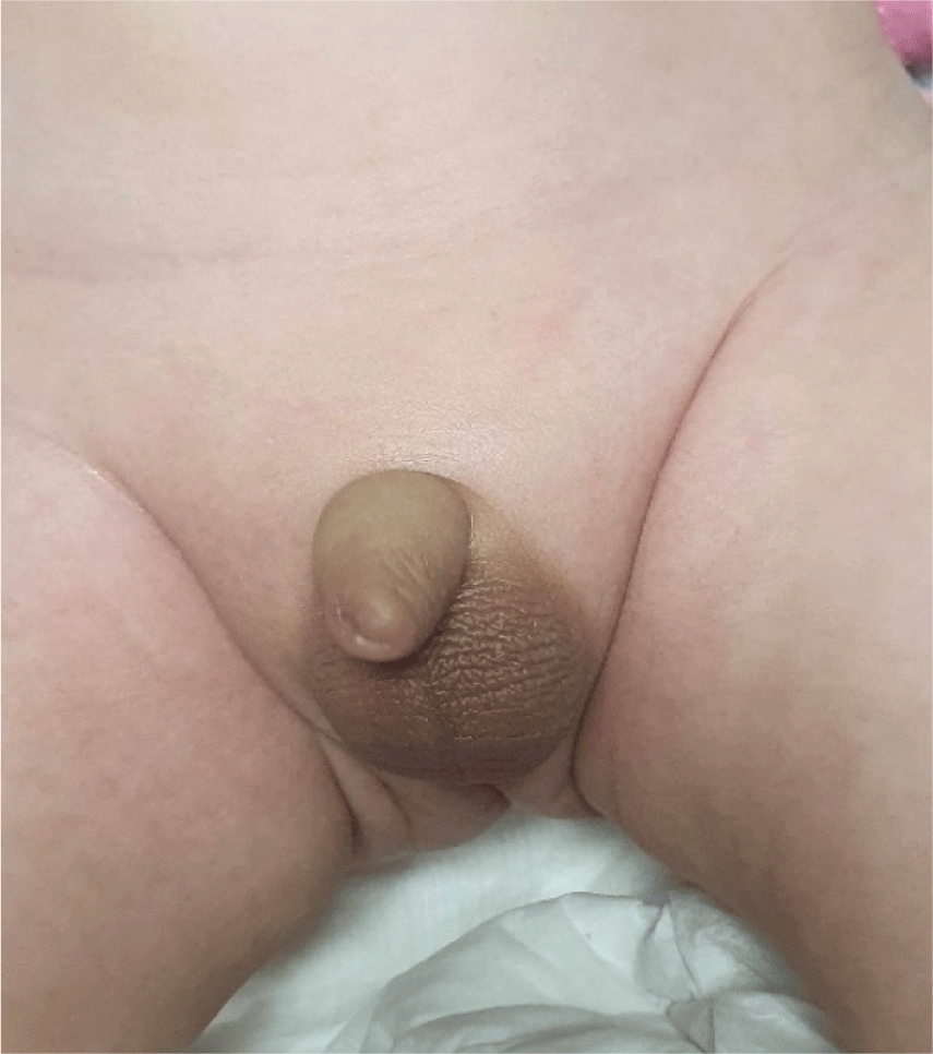
A 3-month-old male infant was admitted to Children’s Hospital 1, Ho Chi Minh City, Vietnam with a four-day history of persistent vomiting, occurring 5–6 times per day, accompanied by significantly reduced formula intake (10–20 mL). He was born to a nonconsanguineous Vietnamese couple at 38 weeks of gestation via caesarean section, with a birth weight of 3,300 g (25th–50th percentile). On examination, his weight was 3,200 g (<3rd percentile) and length was 52 cm (<3rd percentile). Clinical signs of dehydration, including dry lips and delayed capillary refill, were observed. Vital signs showed tachycardia and hypotension, but there were no signs of hyperpigmentation or abnormalities in genitalia (Fig. 1).
Laboratory evaluation showed electrolyte imbalances, including severe hyponatremia (112.5 mEq/L), hyperkalaemia (7.54 mEq/L), and metabolic acidosis (pH 7.23, HCO3- 15.6 mmol/L, base excess –11.9 mmol/L). Blood glucose concentration was within normal range (4.62 mmol/L) with normal serum creatinine (43.2 µmol/L). Urinalysis demonstrated a specific gravity of 1.012 and pH of 5.0, with spot urinary sodium and potassium levels of 65 mEq/L and 27 mEq/L, respectively, indicative of significant renal salt wasting. The hormonal tests, including plasma renin, aldosterone, cortisol, ACTH, and 17-hydroxyprogesterone, were sent to the Medic Medical Center in Ho Chi Minh City, Vietnam, for analysis. Hormonal assessment revealed elevated plasma renin activity (500 µUI/mL; normal range 2.8–39.9 µUI/mL) and aldosterone levels (>100 ng/dL; normal range 4.2–20.9 ng/dL), with normal cortisol (31.15 µg/dL) and ACTH (28 pg/mL). 17-Hydroxyprogesterone levels were initially mildly elevated (16.3 ng/mL; normal range: 1.7–4.0 ng/mL). Given the significant salt loss and an elevated 17-hydroxyprogesterone level, CAH (21-hydroxylase deficiency, salt-wasting form) could not be completely ruled out at the time of presentation. Hydrocortisone stress doses were administered, followed by maintenance therapy with physiological doses of hydrocortisone (15 mg/m2/day), synthetic mineralocorticoid (fludrocortisone) 200 µg/day and salt supplement.
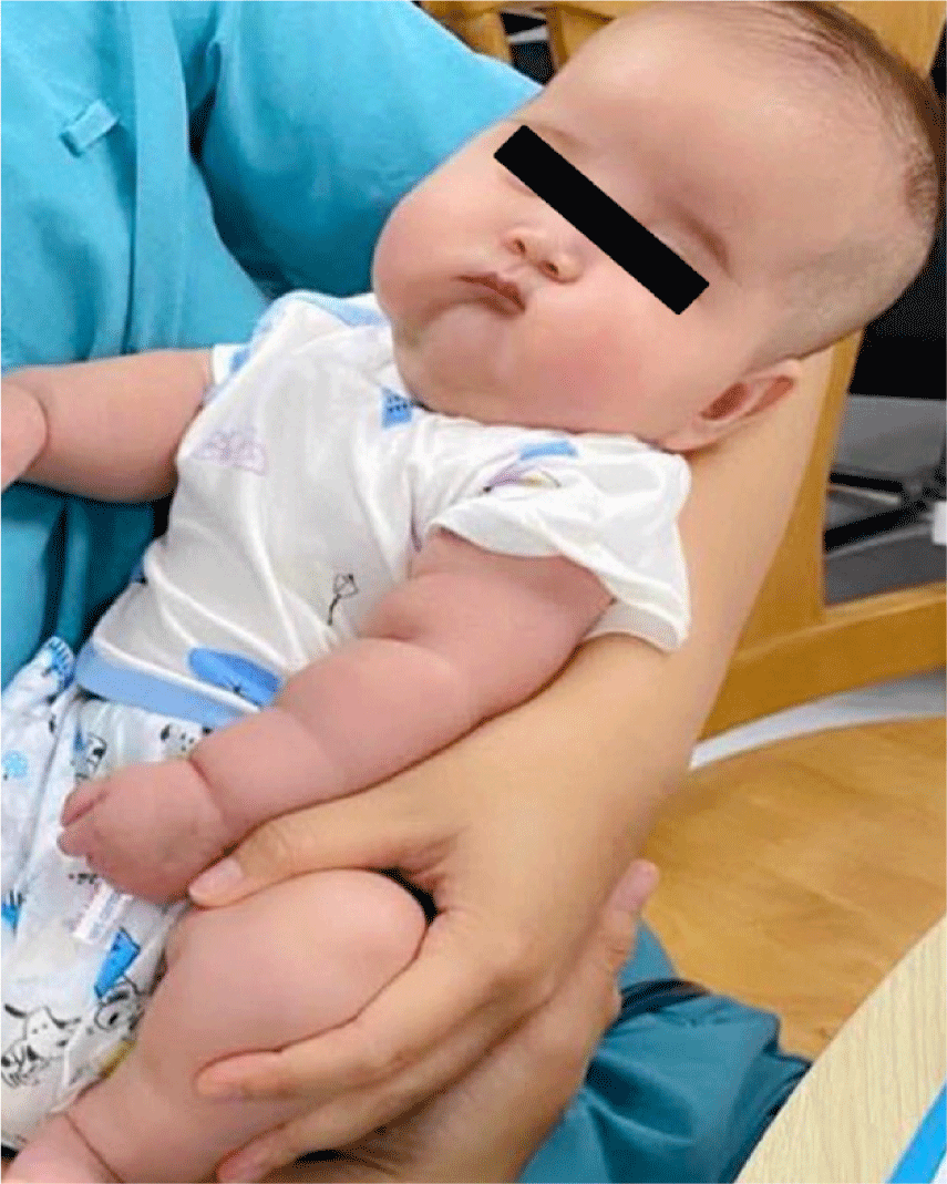
However, one month after discharge, the patient was readmitted with electrolyte disturbances, characterized by hypernatremia and hypokalaemia. On examination, clinical features suggestive of Cushing’s syndrome due to prolonged steroid use were noted (Fig. 2). Repeated hormonal assessment showed persistent elevated plasma renin activity (500 µUI/mL) and aldosterone levels (>100 ng/dL), while ACTH (13 pg/mL) and cortisol (9.3 µg/dL) remained within the respective normal range after discontinuation of hydrocortisone for 48 hours. Additionally, repeated testing of 17-hydroxyprogesterone confirmed normalization (1.42 ng/mL), making CAH a less likely diagnosis. These findings raised concerns about the initial diagnosis, prompting genetic testing for further evaluation.
Under the impression of pseudohypoaldosteronism, genetic testing was indicated. The test was performed in Gene Solution, a genetic testing company in Ho Chi Minh City, Vietnam. A targeted panel for pseudohypoaldosteronism was performed on the following genes: CUL3, HSD11B2, KCNJ5, KLHL3, NR3C2, SCNN1A, SCNN1B, SCNN1G, WNK1, and WNK4.
Genetic testing revealed a heterozygous mutation in the NR3C2 gene (dominant), consistent with PHA1 (Table 1). We gradually titrated the dose of fludrocortisone up to 4 tablets (400 µg/day) to achieve electrolyte stability. The patient subsequently showed significant clinical improvement and normalization of biochemical parameters with appropriate mineralocorticoid therapy and salt supplementation (8 mEq/kg/day).
From 12 months of age, the fludrocortisone dose was gradually tapered without complications. At the 24-month follow-up, the patient remained stable on 50 µg of fludrocortisone per day, with no recurrent hospitalizations, stable electrolyte levels and normal growth.
4. DISCUSSION
PHA1 is a rare disorder with an estimated incidence of approximately 1 in 47,000 live births, with autosomal dominant (AD) PHA1 occurring in 1 in 66,000 [4]. Diagnosing PHA1 can be challenging due to its clinical overlap with other salt-wasting disorders, especially CAH [2,4]. Both conditions often present with symptoms such as hyponatremia, hyperkalaemia, and metabolic acidosis in infancy. CAH is typically associated with abnormal cortisol and mineralocorticoid synthesis, whereas PHA1 is characterized by marked elevations in plasma renin and aldosterone levels [1]. However, laboratory challenges can further complicate the diagnosis of PHA1. In early infancy, transient aldosterone resistance can occur, leading to the increase of renin and aldosterone levels, which may not obviously indicate the diagnosis of PHA1 [5]. Serial biochemical testing, combined with clinical assessment, is crucial for distinguishing between transient aldosterone resistance and mineralocorticoid receptor dysfunction.
Salt-wasting conditions in infants can be caused by congenital adrenal hypoplasia, CAH, aldosterone deficiency, or PHA. Both CAH and congenital adrenal hypoplasia lead to adrenal insufficiency, characterized by low cortisol, elevated ACTH, and decreased aldosterone. However, a single cortisol measurement may not reliably reflect adrenal function, as cortisol secretion is influenced by stress and physiological variability [5]. Even if cortisol levels appear normal, adrenal insufficiency could not be completely ruled out. Because 17-hydroxyprogesterone was mildly elevated (16.3 ng/mL), the diagnosis of CAH was favoured over congenital adrenal hypoplasia. The presence of salt-wasting and suspected CAH led to hydrocortisone treatment, which was later found to be unnecessary. Aldosterone resistance was suspected as the patient exhibited remarkedly elevated aldosterone levels accompanied by electrolyte imbalance. Although PHA was considered during the first hospital admission, aldosterone resistance in neonates and infants can lead to transient elevations in aldosterone levels during the first few months of life [4]. Thus, an increased aldosterone level alone was insufficient to confirm the diagnosis at that time. In the absence of an ACTH stimulation test, due to unavailability of the required medication, diagnosing the cause of salt-wasting remains extremely challenging, and repeated testing was crucial to confirm the diagnosis and exclude CAH. Genetic testing is costly and not covered by national health insurance, so it was not initially advised for the patient.
In this case, repeat testing of 17-hydroxyprogesterone revealed a normalized level of 1.42 ng/mL, effectively ruling out CAH as the leading differential diagnosis. The persistence of electrolyte imbalances despite glucocorticoid therapy, along with elevated renin and aldosterone, prompted genetic testing, which confirmed PHA1 (heterozygous mutation in the NR3C2 gene). Due to cost limitations and the goal of practicing cost-effective medicine, we chose to focus on a targeted panel related to pseudohypoaldosteronism, which helped reduce the patient’s genetic testing expenses. If no mutation is detected in this panel, we will discuss expanding the test to include a broader range of genes.
The condition results from inactivation mutations in the NR3C2 gene, which encodes the mineralocorticoid receptor primarily expressed in the kidney [6]. This leads to aldosterone resistance in the kidney. The disorder typically follows an AD inheritance pattern, though sporadic cases with de novo mutations have been documented [6–8]. In our patient, genetic testing of the parents was declined, preventing confirmation of whether the mutation was inherited or occurred de novo.
Management of PHA1 includes salt supplementation and mineralocorticoid therapy. The dose of fludrocortisone may be titrated up to 500 µg/day (5 tablets) to maintain electrolyte balance [1]. The delay in definitive diagnosis highlights the challenges of differentiating these conditions in clinical practice, especially in settings where genetic testing is not always available, and results often require 3–4 weeks to finalize. Amir Babiker et al. also reported four cases of PHA that experienced delayed diagnosis due to clinical presentations resembling CAH, which is more commonly encountered in clinical practice [2]. Therefore, clinical judgment and repeated biochemical evaluations are essential for timely diagnosis and management. Clinicians should remain cautious in interpreting hormonal results in the neonatal/infant period and consider the possibility of transient aldosterone resistance before confirming a diagnosis of PHA1. The clinical overlap between these conditions can result in unnecessary glucocorticoid treatment, as observed in our patient, and such misdiagnoses are not uncommon in clinical practice. This case provided valuable insights into the real-world challenges of distinguishing PHA1 from CAH, two conditions with overlap clinical and biochemical features. The initial misdiagnosis of PHA1 as CAH led to unwarranted glucocorticoid therapy, highlighting the importance of serial biochemical assessments and careful interpretation of hormonal results in neonates and infants.
A notable aspect of PHA1 is the progressive improvement of aldosterone resistance with age. This natural course can reduce the reliance on high-dose mineralocorticoids and sodium supplementation over time [4,7]. This requires careful tapering of medications to avoid destabilizing electrolyte balance. Clinicians must remain vigilant for signs of hyperkalaemia or hyponatremia during this transition. At the 24-month follow-up, our patient demonstrated improved aldosterone resistance, remaining stable on 50 µg of fludrocortisone per day, with no recurrent hospitalizations.
5. CONCLUSION
In conclusion, this case illustrated the diagnostic and management complexities associated with PHA1. Early differentiation from CAH and other salt-wasting disorders is critical to prevent complications and guide appropriate therapy. Comprehensive hormonal and genetic testing, combined with individualized management, is key to optimizing patient outcomes.