1. INTRODUCTION
Managing a difficult airway in patients with severe spinal deformity poses a significant challenge in clinical practice. Ankylosing spondylitis (AS) is a chronic autoinflammatory disease that primarily affects the axial skeleton and sacroiliac joints, leading to inflammation, pain, stiffness, and progressive spinal fusion, often resulting in the characteristic “bamboo spine” appearance. It predominantly affects males aged 20 to 30 and is strongly associated with the HLA-B27 gene, environmental factors, and immune dysregulation. Chronic inflammation damages ligaments and cartilage, leading to ossification and joint fusion, which reduces spinal mobility and causes fixed deformities such as cervical flexion, restricted mouth opening, and an inability to assume the supine position [1,2]. The anesthesiologist’s role is to identify predictive criteria for difficult intubation, such as a high Mallampati score, restricted mouth opening, limited cervical spine extension, and a shortened sternomental distance. In AS patients, a thorough preoperative physical examination is crucial to assess mechanical limitations in cervical mobility, which represented the primary obstacle in airway management. Various techniques have been proposed in the literature as alternatives to standard intubation to ensure airway control in AS patients. Fiberoptic bronchoscopy is widely regarded as the gold standard in managing anticipated difficult airways, especially in patients with AS. The effectiveness of this method features several key mechanisms including direct visualization of the glottic opening, enabling accurate and atraumatic intubation, and maintenance of spontaneous ventilation throughout the procedure, which reduce the risk of hypoxemia and airway collapse. Also, the flexible structure allows navigation through restricted or distorted anatomical spaces, such as limited cervical mobility or severe kyphosis [1,3].
In this report, we present a patient with AS and severe spinal rigidity who underwent spinal tumor resection and was successfully intubated under flexible fiberoptic bronchoscopy guidance.
2. CASE PRESENTATION
This case report was prepared in compliance with the CARE guidelines to ensure accuracy, transparency, and comprehensiveness of the clinical information provided [4].
A 63-year-old male presented with lower back pain radiating to the right lower extremity, progressive bilateral lower limb weakness, and urinary dysfunction. His medical history was notable for untreated hypertension, and type II diabetes mellitus managed with gliclazide and metformin. He was diagnosed with AS at the age of 15 without receiving disease-specific therapy, resulting in a fixed thoracolumbar kyphotic deformity measuring approximately 60 degrees. There was no relevant family history. Regarding patient psychosocial status, he lived independently, though the spinal condition significantly limited his mobility. His surgical history included posterior spinal fusion for spinal deformity correction at age 15, and bilateral total hip arthroplasty at age 45, both performed under general anesthesia with documented difficult intubations. The patient was admitted to the Department of Neurosurgery at University Medical Center Ho Chi Minh City in Vietnam for scheduled spinal cord tumor resection.
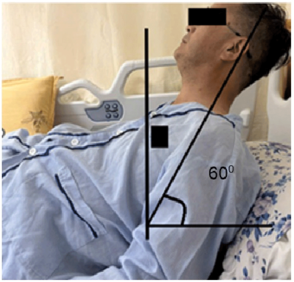
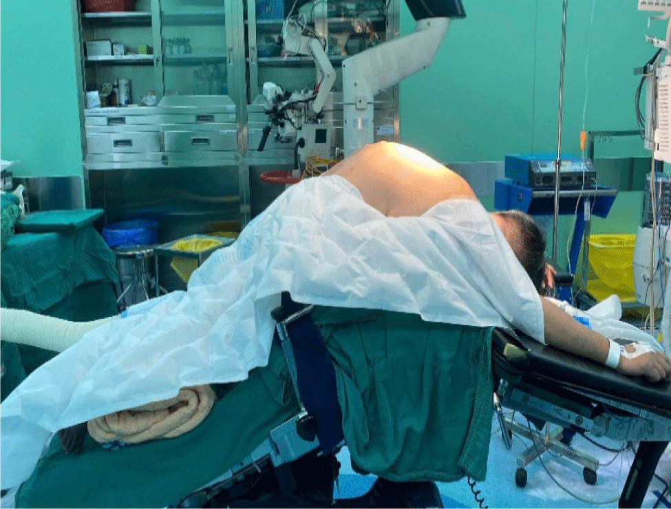
The patient was in no acute distress, with kyphoscoliosis of the spine and significant bilateral lower limb deficits, including positive a Lasegue’s sign, superficial sensory disturbances, and muscle strength of 3/5. The patient presented a fixed 60-degree forward flexion of the spine and complete cervical ankylosis due to long-standing AS. He could only sleep in the lateral decubitus position. When asked to lie supine, he felt uncomfortable and unsafe (Fig. 1).
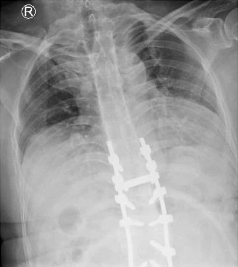
Chest X-ray revealed spinal deformity and ankylosing inflammation involving the cervicothoracic and thoracolumbar spine (Fig. 2).
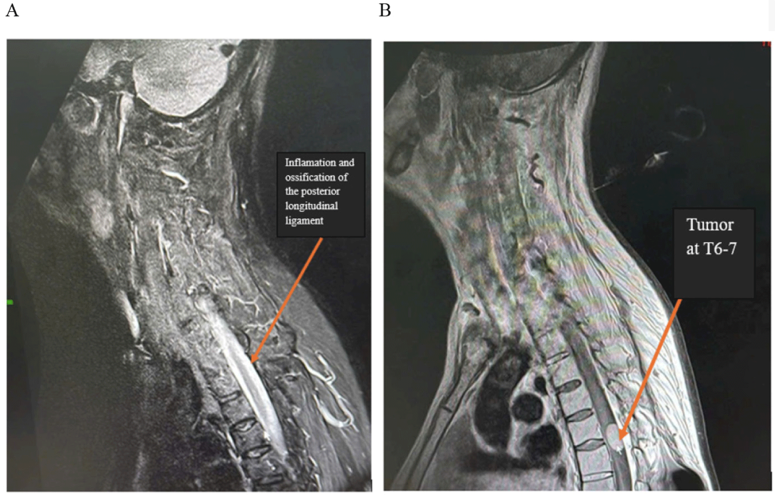
Magnetic resonance imaging (MRI) revealed an intradural extramedullary tumor at the T6–T7 level, 1×2×1.2 cm in size and the ossification of the posterior longitudinal ligament at the cervicothoracic junction and previous spinal instrumentation in the lower thoracic and lumbar regions. The test also revealed hallmark features of AS, accounting for the patient’s cervical rigidity over the years (Fig. 3).
Complete blood test and all biochemical tests, including liver and kidney function, were within normal limits.
Pre-anesthetic evaluation classified the patient as ASA Physical Status III. Although the patient demonstrated a mouth opening of approximately three finger breadths, airway assessment revealed Mallampati class IV. The thyromental distance was approximately one finger breadth, and the sternomental distance was about 1 cm. The cervical and upper thoracic vertebrae were fused, resulting in a fixed forward-flexed posture with the spine bent at approximately 60 degrees, which had persisted for over 40 years. All these findings indicated a challenging airway management scenario.
Preoperative steps included patient identity verification, oxygen supplementation via nasal cannula at 3 L/min and establishing a 20G intravenous line.
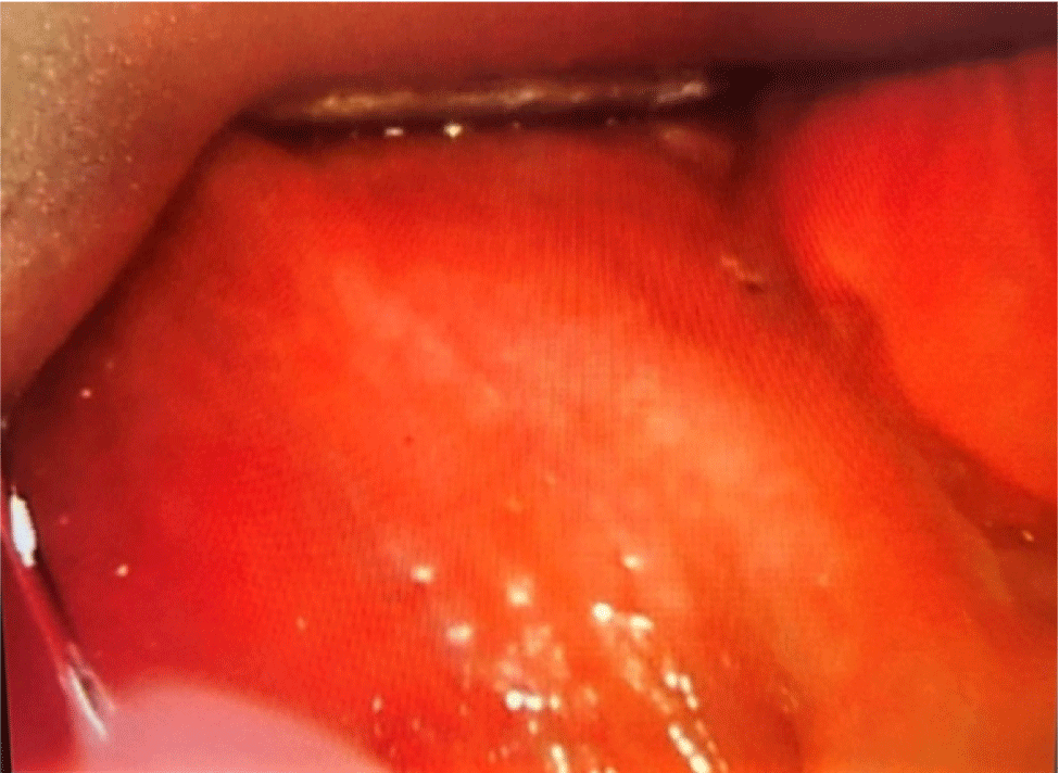
Premedication was administered with fentanyl 50 mcg, and 10% lidocaine spray (eight puffs) was applied to the oropharynx to facilitate airway examination. A C-MAC video laryngoscope was used to assess the airway in both supine and lateral positions, revealing a Cormack-Lehane grade IV view (Fig. 4).
The patient had a 60-degree fixed spinal flexion and complete cervical ankylosis. Attaining an appropriate position for intubation was a challenge in this case due to rigid curvature of the ankylosed spinal column. To prevent neurological injury to the spinal cord and preserve spinal cord function, the prime concerns were minimizing movement during intubation and attaining appropriate position [2].
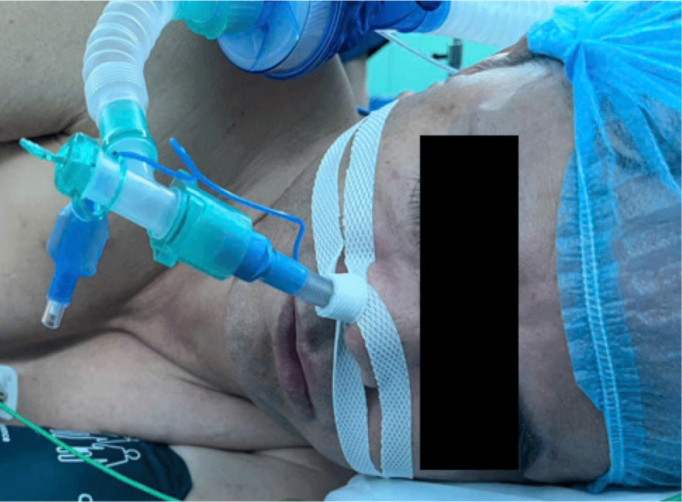
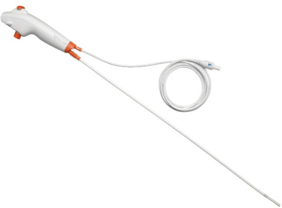
Upon explanation, the patient refused to lie supine. Furthermore, due to the severe forward flexion, the bronchoscopist would have had to stand on a very high platform, increasing the risk of falling. In addition, the patient was at risk of both difficult airway and difficult ventilation. To ensure safety and effective airway control, we decided to perform awake intubation in the left lateral decubitus position. The procedure was performed by an experienced bronchoscopist via the right nostril under the guidance of a flexible fiberoptic bronchoscope (Ambu® ascopeTM, Ambu A/S, Ballerup, Denmark). The patient was lightly sedated using target-controlled infusion (TCI) with propofol at 1 mcg/mL under the Schnider model. Lidocaine 2% 5 mL was used to topicalize the vocal cord, and oxygen was administered via a single-nostril nasal cannula at 5 L/min through the left nostril (Fig. 5).
The larynx, trachea, carina, and both main bronchi appeared normal with intact, mobile vocal cords and no signs of mass or ulceration. Before passing the vocal cords, the patient received an additional fentanyl 100 mcg, propofol 150 mg, and rocuronium 50 mg. A 7.0 mm reinforced endotracheal tube was successfully inserted and secured with the 28 cm mark at the right nostril. The procedure was completed safely in 10 minutes and the patient exhibited stable vital signs without distress throughout (Figs. 6 and 7).
Given the patient’s rigid kyphosis, prone positioning demanded tailored adjustments and teamwork to ensure stability and comfort throughout the surgery. The placement of the endotracheal tube was reverified through auscultation after positioning, ensuring it was correctly positioned in the trachea (Fig. 8).
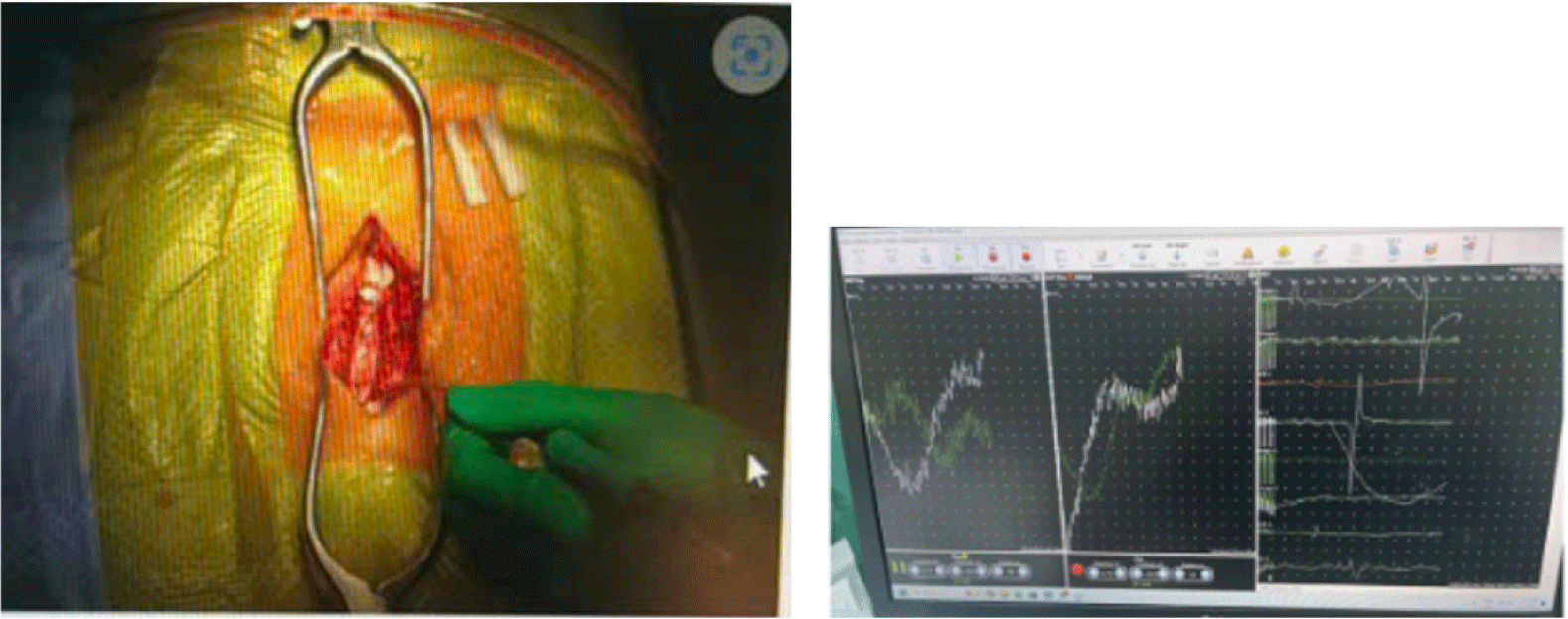
The patient was monitored with electrophysiological monitoring to track the surgical procedures to minimize neurological risks. Throughout the surgery, the patient’s vital signs remained stable, and there was no irregularity regarding the respiratory parameters displayed on the anesthetic machine screen (Fig. 9).
Neuromuscular blockade was reversed with 200 mg sugammadex, and the endotracheal tube was removed when the patient was fully awake and breathing spontaneously with train-of-four ratio >90%. The patient was then placed on nasal oxygen at 3 L/min for 4 hours postoperatively.
The patient was monitored in the post-anesthesia care unit for 4 hours and then transferred to the neurosurgical intensive care unit. Upon arrival, the patient was alert with stable vital signs. There were no symptoms of hoarseness or sore throat. After stable treatment in the neurosurgery ward, the patient was discharged on postoperative day 10.
3. DISCUSSION
In AS, endotracheal intubation is challenging due to limited neck movement from cervical spine fusion, restricted mouth opening from temporomandibular joint involvement, and altered airway anatomy caused by fixed cervical kyphosis. Neurological risks from spinal cord or nerve root compression require careful airway management to avoid further injury. Thus, advanced intubation techniques using flexible fiberoptic scopes, video laryngoscopes, or awake intubation are often necessary to secure the airway safely while maintaining cervical spine stability. Preoperative airway assessment and preparation are crucial to anticipating difficulties and optimizing patient safety [5].
Kotekar described a 50-year-old man with severe AS, a rigid cervical spine, and restricted mouth opening due to temporomandibular joint ankylosis, who was scheduled for hip replacement surgery. The patient was positioned supine with pillow support, and intubation was attempted via conventional direct laryngoscopy following anesthesia induction. Due to poor visualization, three attempts were required, with success only achieved using a specially curved endotracheal tube. Despite maintaining adequate oxygenation, this approach posed risks of airway trauma and hypoxia [6]. In contrast, our case employed awake nasal fiberoptic intubation maintaining spontaneous respiration and visual control throughout. This strategy minimized the risk of airway injury, hypoxemia, and hemodynamic instability. The comparison underscores the superiority of fiberoptic-guided awake techniques in preserving patient safety, particularly in anatomically distorted and high-risk airway scenarios like advanced AS.
A similar reported case by Rebai et al. was also utilized to awake fiberoptic intubation for a patient with severe cervicothoracic kyphosis. Despite a severely compromised airway anatomy, successful awake nasal fiberoptic intubation was achieved in the supine position with meticulous padding and sedation using remifentanil. However, the patient experienced a fatal pulmonary embolism postoperatively, unrelated to the airway technique [7]. In our case, the patient’s longstanding spinal deformity not only restricted anatomical positioning but also induced psychological discomfort in the supine posture. Recognizing this, we selected the lateral decubitus position for awake fiberoptic intubation, allowing for both airway access and emotional reassurance. The comparison underscores that while both cases utilized fiberoptic guided techniques, tailoring positioning to individual anatomical and psychological needs may further enhance safety and patient centered care in difficult airway management.
In Vietnam, with limited healthcare resources, the cost and technical requirements of fiberoptic intubation can hinder routine use. Thus, a preoperative Cormack-Lehane assessment under light sedation can help determine whether flexible bronchoscopy is essential or if conventional methods may suffice. Therefore, in this case, we performed a Cormack-Lehane assessment under mild sedation to devise the most effective plan for the patient. If the Cormack-Lehan score is accepted (I–II), indicating that intubation can be performed, then a flexible endoscope is not necessary. However, in this case, a Cormack-Lehan score of IV indicates that intubation through the C-MAC device is not possible, so a flexible endoscope needs to be used immediately. Centers without access to a flexible endoscope can assess the Cormack-Lehan score under light anesthesia initially to make the first decision. If the Cormack-Lehan score is good enough for intubation, anesthesia and surgery can proceed immediately. However, if the Comack-Lehan score is IV and intubation is difficult (as in our case), transferring the patient to a hospital with a flexible endoscope will allow for safer anesthesia and surgery. During the initial pre-anesthetic evaluation, the patient expressed significant discomfort with the supine position, likely due to his longstanding spinal deformity. After a multidisciplinary team meeting, we opted for awake fiberoptic intubation in the left lateral decubitus position which is uncommon but contextually appropriate choice that ensured both patient comfort and effective airway access. The patient remained comfortable in the familiar lateral position under light sedation with TCI propofol throughout the procedure. This approach differs from the Difficult Airway Society (DAS) guideline on awake tracheal intubation, but it is essential to individualize patient care and consider surrounding clinical factors to select the most appropriate strategy (Table 1) [8].
Thanks to the real-time visualization provided by the flexible endoscope’s camera and the expertise of the endoscopist, we were able to precisely control the scope’s position and successfully perform the endotracheal intubation. Although our experience with this case was encouraging, we acknowledged that further studies are essential to validate the generalizability of this approach in diverse clinical settings.
4. CONCLUSION
Management of difficult airway in patients with severe spinal deformity requires thorough pre-anesthetic assessment and an appropriate intubation strategy. The use of flexible fiberoptic bronchoscopy and awake intubation is a safe and effective choice in cases of fixed cervical spine and inability to lie supine. Thorough preparation, individualized patient planning, and multidisciplinary collaboration are essential especially within limited-resource healthcare systems. These are key factors for successful airway management in patients with spinal deformities.


